Your eyes have a clock sensor, and it is tuned to blue
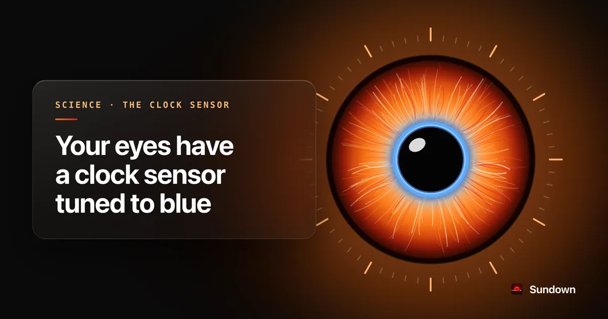
Besides the rods and cones you see with, your retina has a third kind of light sensor. These cells contain a pigment called melanopsin, their main job is to send light information to the brain’s master clock rather than to build images, and they respond most strongly to blue light. That one fact explains why the colour and the brightness of light in the evening matter to your body clock, and it is why Sundown takes the blue out of your Mac’s screen at night.

By Nikki NeislerCo-founder, Sundown · Last reviewed
Key findings
- The retina contains ganglion cells that sense light on their own and report it to the brain’s master clock, separately from the rods and cones used for sight (Berson et al., 2002; Hattar et al., 2002).
- Their pigment, melanopsin, turned up only in the eye when researchers surveyed 26 sites in the human body (Provencio et al., 2000).
- Melatonin suppression is most sensitive to blue light: the most potent band in one study was 446 to 477 nm (Brainard et al., 2001), and blue light shifted the body clock two-fold more than green light of equal photon density (Lockley et al., 2003).
- Bright light still suppressed melatonin in three blind patients with no conscious perception of light (Czeisler et al., 1995), and in a man with no working rods or cones, blue light suppressed melatonin, reset the clock and raised alertness (Zaidi et al., 2007).
- The cones still contribute, mostly at the start of a light exposure and at low light levels. Melanopsin is the main driver in long, bright exposures (Gooley et al., 2010).
- Across 19 lab studies, light measured by how strongly it drives melanopsin was the best available predictor of the clock’s response (Brown, 2020), and ordinary photopic lux is the wrong unit for it (Lockley et al., 2003).
Sundown, tested
Tested by Dr. Alexander Wunsch
Sundown eliminates blue light.
Dr. Alexander Wunsch, a German physician who specialises in light and health, measured a Mac screen across the whole blue band, 380 to 500 nm, with Sundown off and at deep warmth. At deep warmth, the blue is gone.
Sundown removes screen flicker.
Flicker-free mode holds the backlight at full power and does the dimming in software. Jack Richards, who makes Sundown, put the screen on an oscilloscope to check. No flicker.
What is melanopsin?
Melanopsin is a light-sensitive protein made by a small group of cells in the retina called intrinsically photosensitive retinal ganglion cells, usually shortened to ipRGCs. Most retinal ganglion cells only pass on signals that start in the rods and cones. These ones can also detect light by themselves, and the main place they send that information is the suprachiasmatic nucleus, the cluster of neurons in the hypothalamus that keeps the body’s 24-hour time (Berson et al., 2002; Hattar et al., 2002).
When the human gene was first described, the team surveyed 26 sites in the body and found melanopsin expressed only in the eye. Inside the retina it sat in the ganglion and amacrine cell layers, not in the photoreceptors that start vision, and the authors suggested from that location alone that it might handle jobs like setting circadian rhythms and suppressing melatonin rather than forming images (Provencio et al., 2000). The experiments that followed bore that guess out.
How scientists found a third light sensor
The clue came from blind people. In 1995 a team tested 11 patients who had no conscious perception of light at all, exposing them to bright light and measuring their melatonin. In three of them, melatonin fell about as much as it did in sighted volunteers, and when two of those three were tested again with their eyes covered, it did not fall. Those three slept without trouble and kept a normal daily rhythm. Most of the others had no melatonin response and a history of insomnia (Czeisler et al., 1995). Something in those three patients’ eyes was still telling the clock about light, even though the parts of the eye that build pictures no longer worked.
The gene arrived in 2000 (Provencio et al., 2000). Two years later, two papers in Science closed the loop. One showed that the retinal ganglion cells wired to the master clock respond to light directly, even when all input from the rods and cones is blocked, and that their sensitivity, colour tuning and slow response matched those of the clock’s light-setting mechanism (Berson et al., 2002). The other showed that every ganglion cell with that ability carried melanopsin, and traced their wiring in mice to the master clock and to the brain areas that control the pupil (Hattar et al., 2002).
Knockout mice then showed what the sensor does. Mice bred without melanopsin still kept a daily rhythm, but brief pulses of light reset their clocks far less than in normal mice (Panda et al., 2002). Their pupils also behaved oddly: normal in dim light, yet unable to close fully in bright light, which suggested the melanopsin cells and the rod and cone system share the work between them. The ganglion cells wired to the master clock in those mice were no longer sensitive to light by themselves, although their number, shape and wiring were unchanged (Lucas et al., 2003). In monkeys whose rods and cones had been blocked with drugs, the pupil still responded to light, and the slow, lasting squeeze of the pupil after a light switches off turned out to be driven mainly by the melanopsin cells, in monkeys and in people (Gamlin et al., 2007).
The most direct human evidence came in 2007, from two people who had lost their rods and cones entirely. In one of them, short-wavelength light suppressed melatonin, reset the clock and made him more alert than green light at 555 nm, the colour ordinary daytime vision is most sensitive to. In the other, the pupil was most sensitive at 480 nm, matching the melanopsin cells, and she could correctly report a faint flash at that wavelength but at no other (Zaidi et al., 2007). With no working rods or cones, the only sensor left that could explain those results was the one containing melanopsin.
The discovery, in order
| Year | What was found | Study |
|---|---|---|
| 1995 | Bright light suppresses melatonin in some people with no conscious sight | (Czeisler et al., 1995) |
| 2000 | Melanopsin identified in the human eye, in the inner retina | (Provencio et al., 2000) |
| 2001 | Melatonin suppression is most sensitive to blue light, unlike vision | (Brainard et al., 2001; Thapan et al., 2001) |
| 2002 | Retinal ganglion cells that sense light on their own carry melanopsin and wire to the clock | (Berson et al., 2002; Hattar et al., 2002) |
| 2002 to 2003 | Mice without melanopsin reset their clocks poorly and lose part of the pupil reflex | (Panda et al., 2002; Lucas et al., 2003) |
| 2003 | Blue light shifts the human clock two-fold more than green light | (Lockley et al., 2003) |
| 2007 | In two people without working rods or cones, blue light still reached the clock (him) and the pupil peaked near 480 nm (her) | (Zaidi et al., 2007) |
| 2010 | Cones help early on and in dim light; melanopsin drives long, bright exposures | (Gooley et al., 2010) |
| 2014 to 2020 | Light measured for melanopsin (proposed 2014) proves the best predictor of the clock’s response (tested 2020) | (Lucas et al., 2014; Brown, 2020) |
Why is the clock sensor tuned to blue?
Every light sensor in the eye has a colour it responds to best. Daytime vision, run by the cones, is most sensitive at about 555 nm, in the green and yellow part of the spectrum (Lockley et al., 2003). The response that reaches the body clock is not. In 2001 two independent teams built action spectra for melatonin suppression, testing one narrow band of light at a time in the middle of the night.
In the first, 72 volunteers went through 627 tests, and the most potent band for suppressing melatonin was 446 to 477 nm, which is blue (Brainard et al., 2001). In the second, 22 volunteers went through 215 trials, and their curve showed the same strong short-wavelength sensitivity, quite unlike the curves of the rods or the cones (Thapan et al., 2001). The two groups reached the same answer: the photoreceptor that talks to the clock is tuned towards blue, and it is not one of the receptors we see with.
The next question was whether blue light also shifts the clock more. When volunteers sat under blue light at 460 nm for 6.5 hours, their clocks shifted two-fold more than under green light at 555 nm with the same number of photons, and their melatonin was suppressed about twice as much (Lockley et al., 2003). That is a ratio, and it is the honest way to quote this paper: the study reports the comparison between two colours, not a percentage that would apply to anyone’s bedroom.
The tuning shifts a little with age. The lens filters out more blue light as the eye ages, yet in a small study the older group’s melatonin was suppressed just as much as the younger group’s, and their peak sensitivity moved only slightly, from 484 nm in the young to 494 nm in the older participants (Najjar et al., 2014). The authors suspect the system compensates somehow, and say they do not yet know how.
Do rods and cones matter too?
They do. The melanopsin cells receive signals from the rods and cones as well as making their own, so what reaches the clock is a blend (Gooley et al., 2010).
A 2010 study tested this directly by giving people blue light at 460 nm or green light at 555 nm near the time their melatonin normally rises, at a range of intensities. At the start of the exposure, green light suppressed melatonin as well as blue did, which pointed to the cones helping. As the exposure went on, the green light’s effect faded compared with the blue. The authors concluded that cones contribute a lot at the beginning of a light exposure and at low light levels, while melanopsin is the main driver during long, bright exposures (Gooley et al., 2010). The mouse work had already hinted at the same division of labour (Lucas et al., 2003).
So “only blue light matters” is wrong. Brief or dim light of other colours can reach the clock through the cones. What the 2010 study found is that melanopsin is the main driver during long, bright exposures (Gooley et al., 2010), and melanopsin leans towards blue.
What does the clock sensor control?
The melanopsin cells do more than set the clock. The studies on this page tie them to five separate things your body does with light:
- Clock timing. Light at the wrong time of day shifts the clock later or earlier, and short-wavelength light shifts it more than green light (Lockley et al., 2003; Panda et al., 2002).
- Melatonin. Light at night suppresses it, most strongly in the blue band (Brainard et al., 2001; Thapan et al., 2001).
- The pupil. The lasting constriction after bright light depends mainly on the melanopsin cells (Lucas et al., 2003; Gamlin et al., 2007).
- Alertness. In a man with no working rods or cones, blue light raised alertness more than green light did (Zaidi et al., 2007).
- Rudimentary visual awareness. A woman with no working rods or cones could correctly report a faint flash near 480 nm but not at other wavelengths (Zaidi et al., 2007).
That last point means the same light that suppresses melatonin can also raise alertness (Zaidi et al., 2007). How much ordinary evening light it takes, and how alert it makes people, is the subject of another article in this library, ordinary evening light moves your body clock.
Measuring light the way the clock sees it
Lux, the unit on a light meter, weights light by how bright it looks to daytime vision, so it is dominated by the green and yellow light the cones like best. Once it was clear that a different sensor runs the clock, researchers from across the field proposed a new way to measure light that accounts for every input the clock receives, melanopsin included, and offered simple suggestions for lighting designers and regulators (Lucas et al., 2014).
One measure of this kind is melanopic illuminance. It counts how much light reaches the eye and weights it by how strongly each wavelength drives melanopsin. When one researcher pooled 19 laboratory studies, melanopic illuminance was the best available predictor of how the human circadian system responded across all of them (Brown, 2020). Two lights that look equally bright can differ a lot on this scale, which is why a lamp’s colour and its brightness both matter at night.
What this means for screens at night
A computer screen is a source of light at eye level, often for hours in the evening. When a tablet, an e-reader and a phone were measured, all three gave off light with a strong blue peak (Gringras et al., 2015). Two things lower how much a screen drives the clock sensor: making it warmer, so less of its light falls in the blue band, and making it dimmer, so less light arrives at all.
The trials that tested only the first lever are a useful warning. On an iPad, both of Night Shift’s settings still suppressed melatonin, the low and high colour-temperature settings did not differ significantly, and the authors concluded that changing a screen’s colour without changing its brightness may not be enough (Nagare et al., 2019). The full set of screen studies, including the ones that found no effect, is on the page on screens at night and sleep, and the Night Shift trials have a page of their own.
Sundown is built on exactly this. It warms a Mac’s display down to a deep red, where Night Shift stops at a warm white, and dims it as well, so it works on both the colour and the amount of light. Dr. Alexander Wunsch measured it across the blue band, 380 to 500 nm, the range that takes in melanopsin’s peak, and at deep warmth Sundown eliminates blue light. The method is on how we tested Sundown.
Questions people ask
- Is melanopsin the same as melatonin?
- No. Melanopsin is the light-sensing protein in certain retinal cells. Melatonin is a hormone released at night. Light picked up by the melanopsin cells is one of the main things that suppresses melatonin, which is why the two names come up together (Provencio et al., 2000; Brainard et al., 2001).
- What colour of light suppresses melatonin the most?
- Blue. The most potent band in the largest action-spectrum study was 446 to 477 nm (Brainard et al., 2001). Green light can also suppress melatonin at the start of an exposure, through the cones (Gooley et al., 2010).
- Can light affect the body clock of someone who is blind?
- In some people, yes. Bright light suppressed melatonin in three of 11 patients with no conscious sight, and blue light reset the clock of a man with no working rods or cones (Czeisler et al., 1995; Zaidi et al., 2007).
- Does a warmer screen help?
- A warmer screen gives off less of the light melanopsin responds to, but changing colour without changing brightness may not be enough: in one iPad trial, both Night Shift settings still suppressed melatonin and the warm one did no significantly better than the cool one (Nagare et al., 2019). Warmth and lower brightness together reduce melanopic light the most. Sundown does both, and at deep warmth it eliminates blue light.
- What is an ipRGC?
- An intrinsically photosensitive retinal ganglion cell: a retinal cell that contains melanopsin, detects light by itself and sends that signal to the brain’s master clock and to the areas that control the pupil (Berson et al., 2002; Hattar et al., 2002).
Put the research to work on your Mac
Sundown warms your Mac’s display down to a deep red and dims it after dark. Tested by Dr. Alexander Wunsch, it eliminates blue light. Its flicker-free mode removes screen flicker too.
The research
Every study cited above, with its design, its sample size and what it found, linked to the paper. Each record was read on PubMed or the journal’s site.
- 01
Czeisler CA, Shanahan TL, Klerman EB, et al. (1995). Suppression of melatonin secretion in some blind patients by exposure to bright light. The New England Journal of Medicine.
Controlled clinical trial · n: 11 blind patients, 6 sighted controls
Found: In three blind patients with no conscious perception of light, bright light still lowered melatonin about as much as in sighted people. In the two of them retested with their eyes covered, it did not.
Limits: Small numbers. Most of the blind patients showed no response.
PubMed 7990870 ↗DOI 10.1056/NEJM199501053320102 - 02
Provencio I, Rodriguez IR, Jiang G, et al. (2000). A novel human opsin in the inner retina. The Journal of Neuroscience.
Lab study · n: Human tissue survey; primate and mouse retina
Found: Identified melanopsin, a light-sensitive protein found in the human eye and nowhere else in the body, sitting in the inner retina rather than in the rods and cones.
Limits: Gene and tissue work. It proposed a clock role for melanopsin; it did not test one.
PubMed 10632589 ↗DOI 10.1523/JNEUROSCI.20-02-00600.2000 - 03
Berson DM, Dunn FA, Takao M (2002). Phototransduction by retinal ganglion cells that set the circadian clock. Science.
Lab study · n: Rat retina
Found: Found a third kind of light-sensing cell in the retina that reports light straight to the brain's master clock, separate from the rods and cones you see with.
Limits: Animal tissue. It shows the sensor exists, not what any screen does to people.
PubMed 11834835 ↗DOI 10.1126/science.1067262 - 04
Hattar S, Liao HW, Takao M, Berson DM, Yau KW (2002). Melanopsin-containing retinal ganglion cells: architecture, projections, and intrinsic photosensitivity. Science.
Lab study · n: Rat and mouse retina
Found: Every retinal ganglion cell that responded to light on its own carried melanopsin, and these cells sent their wiring to the brain's master clock.
Limits: Animal tissue.
PubMed 11834834 ↗DOI 10.1126/science.1069609 - 05
Panda S, Sato TK, Castrucci AM, et al. (2002). Melanopsin (Opn4) requirement for normal light-induced circadian phase shifting. Science.
Lab study (knockout mice) · n: Mice
Found: Mice bred without melanopsin still kept a daily rhythm, but brief pulses of light reset their clocks far less than in normal mice.
Limits: Mice. Rods and cones still did part of the job, which is why the clock was weakened rather than blind.
PubMed 12481141 ↗DOI 10.1126/science.1076848 - 06
Lucas RJ, Hattar S, Takao M, et al. (2003). Diminished pupillary light reflex at high irradiances in melanopsin-knockout mice. Science.
Lab study (knockout mice) · n: Mice
Found: Without melanopsin, the pupil reacted normally to dim light but failed to close fully in bright light: the two light systems share the work.
Limits: Mice, and the pupil rather than the clock.
PubMed 12522249 ↗DOI 10.1126/science.1077293 - 07
Gamlin PDR, McDougal DH, Pokorny J, et al. (2007). Human and macaque pupil responses driven by melanopsin-containing retinal ganglion cells. Vision Research.
Lab study · n: Macaques and human volunteers
Found: The pupil's slow, lasting squeeze after a light goes off is driven mainly by the melanopsin cells, in monkeys and in people.
Limits: Pupil responses, not the clock or sleep.
PubMed 17320141 ↗DOI 10.1016/j.visres.2006.12.015 - 08
Zaidi FH, Hull JT, Peirson SN, et al. (2007). Short-wavelength light sensitivity of circadian, pupillary, and visual awareness in humans lacking an outer retina. Current Biology.
Case reports · n: 2 blind patients
Found: Two people with no working rods or cones: in the man, short-wavelength light suppressed melatonin, reset the clock and raised alertness more than 555 nm light; in the woman, pupil sensitivity peaked at 480 nm and she could report a faint 480 nm flash but no other wavelength.
Limits: Two people.
PubMed 18082405 ↗DOI 10.1016/j.cub.2007.11.034 - 09
Brainard GC, Hanifin JP, Greeson JM, et al. (2001). Action spectrum for melatonin regulation in humans: evidence for a novel circadian photoreceptor. The Journal of Neuroscience.
Lab study · n: 72 adults, 627 tests
Found: Mapped which colours of light suppress melatonin at night. The most potent band was 446 to 477 nm, which is blue.
Limits: Single-colour light at 2 to 3:30 in the morning with dilated pupils, not a screen.
PubMed 11487664 ↗DOI 10.1523/JNEUROSCI.21-16-06405.2001 - 10
Thapan K, Arendt J, Skene DJ (2001). An action spectrum for melatonin suppression: evidence for a novel non-rod, non-cone photoreceptor system in humans. The Journal of Physiology.
Lab study · n: 22 adults, 215 trials
Found: An independent team found the same thing: melatonin suppression peaks in the short-wavelength range, very differently from normal vision.
Limits: 30-minute pulses of single-colour light in a lab.
PubMed 11507175 ↗DOI 10.1111/j.1469-7793.2001.t01-1-00261.x - 11
Lockley SW, Brainard GC, Czeisler CA (2003). High sensitivity of the human circadian melatonin rhythm to resetting by short wavelength light. The Journal of Clinical Endocrinology & Metabolism.
Lab study · n: Not stated in abstract
Found: 6.5 hours of blue (460 nm) light shifted the body clock two-fold more than green (555 nm) light of equal photon density, and suppressed about twice as much melatonin.
Limits: Single-colour light for 6.5 hours, not a screen. The paper reports ratios, not percentages.
PubMed 12970330 ↗DOI 10.1210/jc.2003-030570 - 12
Najjar RP, Chiquet C, Teikari P, et al. (2014). Aging of non-visual spectral sensitivity to light in humans: compensatory mechanisms? PLOS ONE.
Lab study · n: 8 older and 5 young adults
Found: Older eyes let through less blue light, yet melatonin suppression was not reduced. The peak sensitivity moved slightly, from 484 nm in the young to 494 nm in the older group.
Limits: Thirteen people, single-colour light.
PubMed 24465738 ↗DOI 10.1371/journal.pone.0085837 - 13
Gooley JJ, Rajaratnam SMW, Brainard GC, et al. (2010). Spectral responses of the human circadian system depend on the irradiance and duration of exposure to light. Science Translational Medicine.
Lab study (dose-response) · n: Not stated in abstract
Found: At the start of a light exposure and at low light levels, cones helped suppress melatonin too. Over longer and brighter exposures, melanopsin took over as the main driver.
Limits: Single-colour blue and green light in a lab.
PubMed 20463367 ↗DOI 10.1126/scitranslmed.3000741 - 14
Lucas RJ, Peirson SN, Berson DM, et al. (2014). Measuring and using light in the melanopsin age. Trends in Neurosciences.
Review (expert group) · n: Review
Found: Researchers across the field proposed measuring light in a way that accounts for every input the eye's non-visual responses receive, melanopsin cells as well as rods and cones, instead of by brightness to vision alone.
Limits: A proposal for how to measure light, not an experiment.
PubMed 24287308 ↗DOI 10.1016/j.tins.2013.10.004 - 15
Brown TM (2020). Melanopic illuminance defines the magnitude of human circadian light responses under a wide range of conditions. Journal of Pineal Research.
Pooled analysis · n: 19 lab studies
Found: Across 19 lab studies, one measure best predicted the clock's response: melanopic illuminance, which counts how much light reaches the clock sensor and how well its colour drives it.
Limits: Built from laboratory studies, not from people's own homes or screens.
PubMed 32248548 ↗DOI 10.1111/jpi.12655 - 16
Gringras P, Middleton B, Skene DJ, Revell VL (2015). Bigger, Brighter, Bluer-Better? Current Light-Emitting Devices: Adverse Sleep Properties and Preventative Strategies. Frontiers in Public Health.
Measurement study · n: 3 devices
Found: A tablet, an e-reader and a phone all gave off light with a strong blue peak. The authors call for a bedtime mode that shifts the screen to yellow and red and also lowers its brightness.
Limits: Measured the devices, not anyone's sleep.
PubMed 26528465 ↗DOI 10.3389/fpubh.2015.00233 - 17
Nagare R, Plitnick B, Figueiro MG (2019). Does the iPad Night Shift mode reduce melatonin suppression? Lighting Research & Technology.
Crossover trial · n: 12 adults (full text)
Found: Both Night Shift settings, warm and cool, still suppressed melatonin, and the two did not differ significantly. The authors conclude that changing a screen's colour without changing its brightness may not be enough.
Limits: Twelve people, an iPad. There was no condition with Night Shift off, so it cannot show whether Night Shift helps compared with off.
PubMed 31191118 ↗DOI 10.1177/1477153517748189
How to cite this page
Neisler, N. (2026). Your eyes have a clock sensor, and it is tuned to blue. Sundown. https://trysundown.com/science/melanopsin-and-blue-light (last reviewed 11 October 2026).
Found a study we missed, a link that has rotted or a sentence that overstates a paper? Write to support@trysundown.com and we will correct it and update the review date.
Notes on the research
- Most of the human experiments used single-colour light in a laboratory, often in the middle of the night and sometimes with dilated pupils (Brainard et al., 2001; Thapan et al., 2001; Lockley et al., 2003). Screens give off a broad mix of wavelengths at a lower intensity.
- Several key findings come from animal tissue or knockout mice (Berson et al., 2002; Hattar et al., 2002; Panda et al., 2002; Lucas et al., 2003). They show how the system is built, not what a given amount of light does to a person.
- The blind-patient studies are small: three responders in one study and two people in the other (Czeisler et al., 1995; Zaidi et al., 2007).
- Cones contribute too, especially in dim or brief light, so blue light is not the only route to the clock (Gooley et al., 2010). People also differ, and age shifts the peak slightly (Najjar et al., 2014).
- Sundown is a display utility, not a medical device. It does not diagnose or treat any condition.